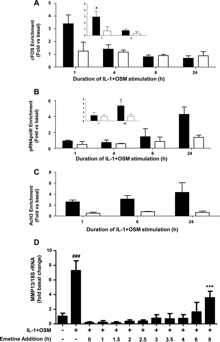
FIGURE 2.
ChIP analyses of the MMP13 proximal promoter. Human chondrocytes were treated with IL-1 (0.05 ng/ml) in combination with OSM (10 ng/ml) for the indicated durations. Cells were then subject to DNA-protein cross-linking, lysis, and DNA shearing. Immunoprecipitation for cFOS (A), pRNA Pol II (B), or AcH3 (C) was followed by isolation of complexed genomic DNA and subsequent real-time RT-PCR for the proximal (closed bars) and 3′-UTR (used for normalization; open bars) regions of MMP13 as indicated. Data (mean ± S.D.) are pooled from three (five for inset data) separate chondrocyte populations. Statistical comparisons are: *, p < 0.05 (24 h IL-1 + OSM stimulation versus basal); #, p < 0.05 (1 h IL-1 + OSM stimulation versus basal). D, chondrocytes were treated with IL-1 (0.05 ng/ml) and OSM (10 ng/ml) for 24 h. Emetine (10 μm final concentration) was added at the indicated times after IL-1 + OSM stimulation and real-time RT-PCR performed on extracted RNA relative expression levels of MMP13 mRNA were normalized to 18S rRNA, where ***, p < 0.001 (IL-1 + OSM + emetine versus basal); ###, p < 0.001 (IL-1+OSM versus basal). Data (mean ± S.D., n = 6) are representative of three separate experiments each using chondrocyte cultures from different donors.

