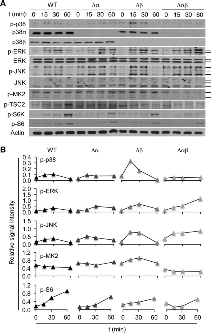
FIGURE 2.

p38α and p38β cooperate to shape TCR-induced intracellular signaling in CD4+ T cells. A and B, naïve CD4+ T cells from the indicated mice were left unstimulated or stimulated with anti-CD3 and anti-CD28. Whole cell lysates were prepared after the indicated durations of stimulation and analyzed by immunoblotting (A). Solid bars on the right indicate bands corresponding to multiple protein isoforms detected by the antibodies. p-, phosphorylated. Immunoblot signals were quantified by densitometry, and the relative signal intensities (the indicated proteins relative to actin) are shown (B). Data are representative of three experiments with similar results (A and B).
