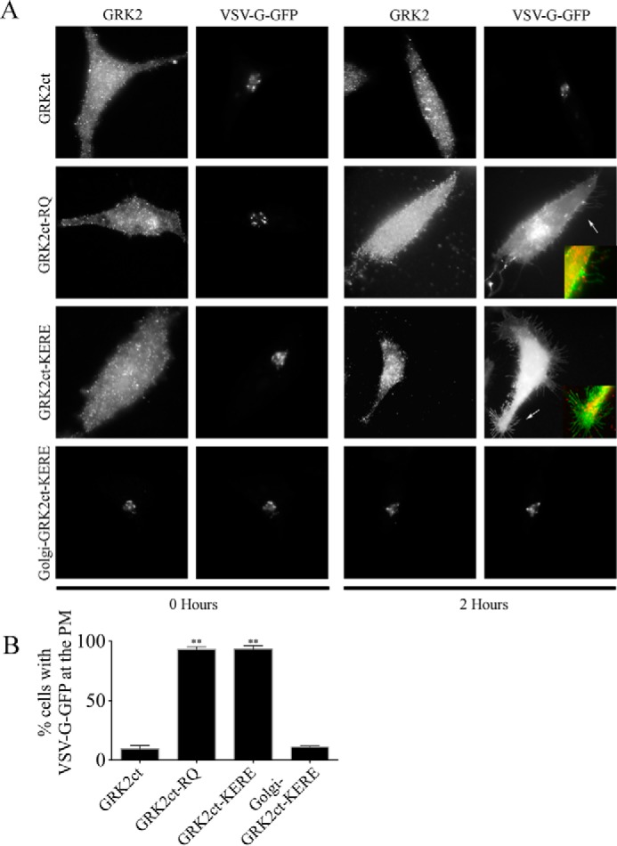
FIGURE 1.

Golgi-GRK2ct-KERE inhibits VSV-G PM transport. HeLa cells were transfected with expression plasmids for temperature-sensitive VSV-G-GFP and GRK2ct, WT or mutant, as indicated. VSV-G-GFP cargo accumulates in the ER, then Golgi, and then transports to the PM when cells are incubated at 39.5, 20, and 32 °C, respectively, as described under “Experimental Procedures.” Cells were fixed at the start of the 32 °C incubation (0 Hours) or 2 h after the shift to 32 °C. A, cells were processed for immunofluorescence microscopy using anti-GRK2 and anti-GFP antibodies to detect the localization of GRK2ct (WT or mutant) and VSV-G-GFP, respectively. Representative images are shown. Inset panels, corresponding to arrows, show dual color images of VSV-G-GFP (green) and GRK2ct-RQ or GRK2ct-KERE (red) to clearly identify PM localization of the VSV-G-GFP cargo. B, percentage of transfected cells with detectable PM localization of VSV-G-GFP after 2 h at 32 °C, as described under “Experimental Procedures.” More than 150 cells were scored for PM localization of VSV-G-GFP in each of n = 5 experiments, and the bars represent the average and S.D. (error bars). Statistical significance compared with WT GRK2ct was tested using an unpaired t test (**, p < 0.0001).
