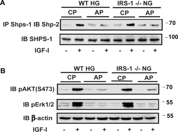
FIGURE 3.

Disruption of SHPS-1 signaling complex formation prevents enhanced IGF-I-dependent MAPK and AKT activation. A, aortic lysates from diabetic wild-type mice (WT HG) and normal IRS-1 knockout mice (IRS-1−/− NG) that had been treated with a control peptide (CP) or a SHPS-1/SHP-2-disrupting peptide (AP) were immunoprecipitated (IP) with an anti-SHPS-1 antibody and immunoblotted (IB) with an anti-SHP-2 antibody. The same amount of each aortic lysate was immunoblotted with an anti-SHPS-1 antibody as a loading control. Scanning densitometry values obtained from of the aortic extracts from three mice in two separate experiments showed a 5.8 ± 0.9-fold increase in wild-type diabetic mice exposed to the control peptide and a 1.6 ± 0.3-fold (p < 0.01) change with the disrupting peptide. The increases were 6.4 ± 0.7-fold and 1.4 ± 0.2-fold (p < 0.01), respectively, in IRS-1−/− mice. B, the aortic extracts were immunoblotted with anti-pAKT (Ser-473) and pErk1/2 antibodies. The blots were reprobed with an anti-β-actin antibody to control for loading. The increases AKT activation were 9.5 ± 1.7-fold and 1.7 ± 0.2-fold (p < 0.001) in wild-type diabetic and 9.1 ± 1.5-fold compared with 2.0 ± 0.3-fold (p < 0.01) in IRS-1−/− mice. The changes in MAPK were 6.0 ± 1.0-fold and 1.2 ± 0.2-fold (p < 0.01) in diabetic wild-type mice and 7.9 ± 1.6-fold compared with 1.3 ± 0.3-fold (p < 0.01) in IRS-1−/− mice.
