Figure 6.
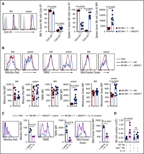
Autophagy and mitophagy are enhanced during GVHD. (A) B6 and B6D2F1 mice were transplanted with B6.CD45.1 BM and CD3+ T cells. The frequency of Cyto-ID+ NK cells in the BM and spleen and the Cyto-ID MFI in the spleen of donor NK cells was quantified. Representative histograms at day 14 posttransplant are shown. Mean ± SEM pooled from 2 independent experiments; n = 8 per group. (B) Levels of MitoSox Red, MitoTracker Green, and TMRE were examined in the donor NK cells from the mice in panel A. MFI values for the 3 mitophagy stains were quantified. Representative histograms at day 14 posttransplant are shown. Mean ± SEM pooled from 2 independent experiments; n = 10 per group. (C) B6.BM and B6.CD3+ T cells was transplanted into lethally irradiated B6D2F1 recipients and treated on day 7 and day 10 with saline or IL-15 complex. Quantitation of MitoSox Red, TMRE, and MitoTracker Green levels in donor NK cells from the BM at day 14. Dotted line indicates level in a nontransplanted B6 mouse. Mean ± SD from 1 experiment; n = 3 per group. (D) Enumeration of donor NK cells in the BM at day 14 posttransplant after BM from B6.NKp46cre+.Atg7WT/WT or B6.NKp46cre+.Atg7fl/fl was transplanted together with B6.CD3+ T cells into lethally irradiated B6D2F1 recipients and treated as in panel C. Mean ± SEM pooled from 2 independent experiments; n = 6-8 per group. Mann-Whitney tests were performed in panels A and B. The unpaired t test with Welch correction was performed in panels C and D. FMO, fluorescence minus one.

