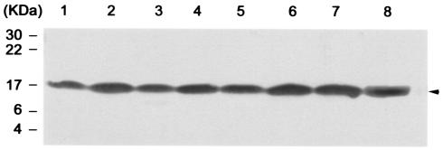
FIG. 3.
Western blot analysis of F1 antigen expressed by Y. pestis KIM5-3001 derivatives. The arrowhead indicates the F1 antigen. Lanes:1, KIM5-4003 (KIM5-3001.1 carrying pDG5); 2, KIM5-4004 (KIM5-3001.1 carrying pIC20R); 3, KIM5-4005 (KIM5-4002 carrying pDG5); 4, KIM5-4006 (KIM5-4002 carrying pIC20R); 5, KIM5-3001 (psaA+ pCD1+); 6, KIM5-3001.1 (psaA mutant pCD1+); 7, KIM5-4001 (psaA+ pCD1−); 8, KIM5-4002 (psaA mutant pCD1−). An equal amount of bacteria was loaded in each lane (see Materials and Methods).

