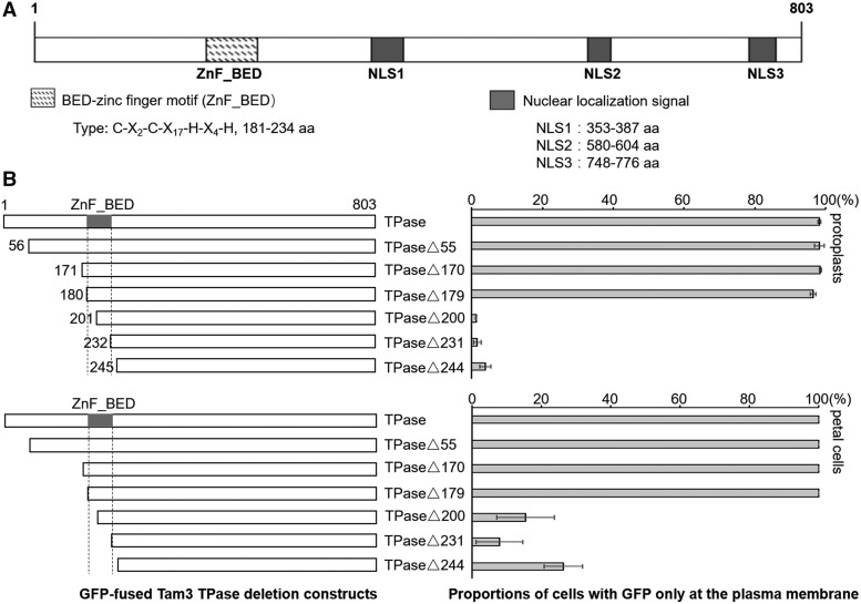
Figure 2.
The Znf-BED motif of Tam3 TPase functions to direct Tam3 TPase to the PM. A, Protein domains of Tam3 TPase. The TPase contains a conserved Znf-BED motif and three experimentally confirmed NLSs. B, Identification of the potential functional domain of Tam3 TPase, which is responsible for directingTam3 TPase to the PM. Left, schematic of the fusion proteins used. Full-length or truncated TPase was fused to the N-terminal region of GFP. Each of these was transformed into protoplasts (top) and petal cells (bottom) of HAM22 to analyze subcellular localization. Numbers in the constructs represent the amino acid position in the Tam3 TPase sequence. Right, two histograms showing the proportions of cells with GFP signal in the PM compared with the total number of cells with green fluorescence (Supplemental Table S1). Data represent means ± sd.

