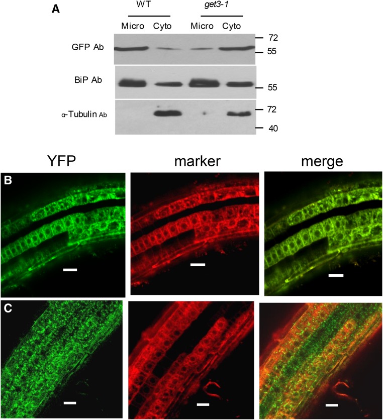
Figure 4.
Subcellular localization of YFP-SYP72 in wild-type and get3-1 backgrounds. A, Wild-type and get-3-1 seedlings from lines stably expressing YFP-SYP72 were fractionated into microsomal and cytoplasmic fractions. Immunoblots of fractionated and total extracts were probed with anti-GFP and anti-BiP antibodies. BiP was used as an ER marker. B and C, Confocal microscopy of roots from seedlings coexpressing YFP-SYP72 and the CDC-960 mCherry ER marker in a wild-type background (B) and a get-3 background (C). D, Roots from seedlings expressing YFP-SYP72 in a wild-type background counter stained with propidium iodide. Bar = 50 µm.

