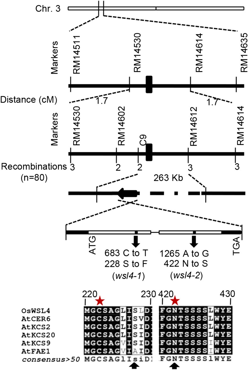
Figure 3.
Mapping-based cloning and structure of WSL4. The schematic diagram depicts the exon (empty box), intron (black line), and untranslated region (solid black box) of WSL4. Amino acid sequence depicting the changes due to point mutations in the two mutants (wsl4-1 and wsl4-2; black arrows). Asterisks indicate the conserved active-site residues in Arabidopsis KCSs.

