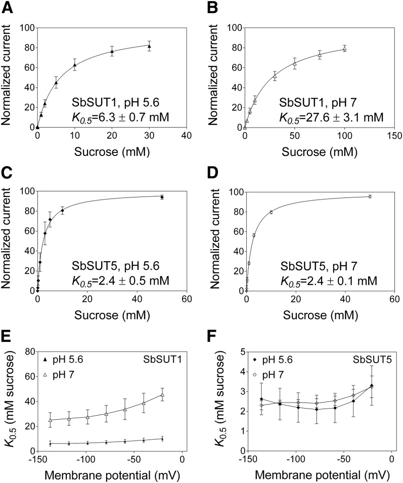
Figure 2.
Voltage and pH dependences of the affinities of SbSUT1 and SbSUT5 for Suc. Transporters were expressed in X. laevis oocytes and recordings made by two-electrode voltage clamping. A to D, Concentration-dependent Suc transport. A, SbSUT1 at pH 5.6. B, SbSUT1 at pH 7. C, SbSUT5 at pH 5.6. D, SbSUT5 at pH 7. Currents were recorded under voltage-clamped conditions at a membrane potential of −117 mV. A Michaelis-Menten curve was fitted to each data set, which was then normalized to Vmax and plotted against the substrate concentration. E and F, Voltage dependence of SUT affinity for Suc. E, SbSUT1 Suc affinities at membrane potentials from −137 mV to −20 mV. F, SbSUT5 Suc affinities at membrane potentials from −137 mV to −20 mV. Mean ± sd (vertical bars) of three to five oocytes.

