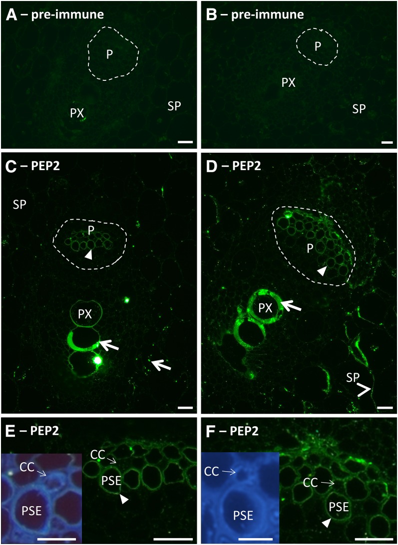
Figure 5.
Immunolocalization of SbSUTs with PEP2 antiserum in apoplasmic phloem unloading zones of elongating Internode 10. A, C, and E, Meristematic zone. A, Preimmune with secondary antibody control. C, Vascular bundle with the phloem outlined by a broken white line. E, Higher magnification of phloem shown in C; inset shows UV autofluorescent image of E. B, D, and F, Elongating zone. B, Preimmune with secondary antibody control. D, Vascular bundle with the phloem outlined by broken white line. F, Higher magnification of phloem shown in D; inset shows UV autofluorescent image of F. Immunolabeling by PEP2 antiserum was present on plasma membranes of PSEs in C to F (darts) and SP cells (arrowheads) in D. CCs did not appear to be labeled. Nonspecific labeling of protoxylem element walls in C and D, and starch grains in C (arrows). Enlarged preimmune control images shown in Supplemental Figure S6. Sections were viewed under the fluorescein isothiocyanate filter (excitation, 450 to 490 nm; emission, >515 nm) and the UV filter (excitation, 365 nm; emission, >420 nm). P, Phloem; PX, protoxylem element. Bars = 20 µm; inset bar = 10 µm.

