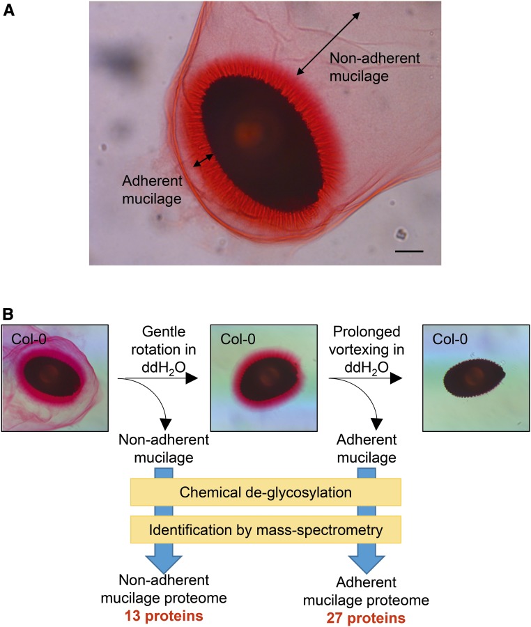
Figure 1.
Strategy to isolate and identify seed coat mucilage proteins. A, Columbia-0 (Col-0) seed coat mucilage stained with Ruthenium Red. Double-headed arrows depict the two mucilage layers. Bar = 100 µm. B, Schematic depiction of the extraction and identification of mucilage proteins. The nonadherent mucilage and adherent mucilage were extracted sequentially. Proteins in each mucilage layer were identified by mass spectrometry (MS) after chemical deglycosylation and trypsin digestion. ddH2O, Distilled, deionized water.

