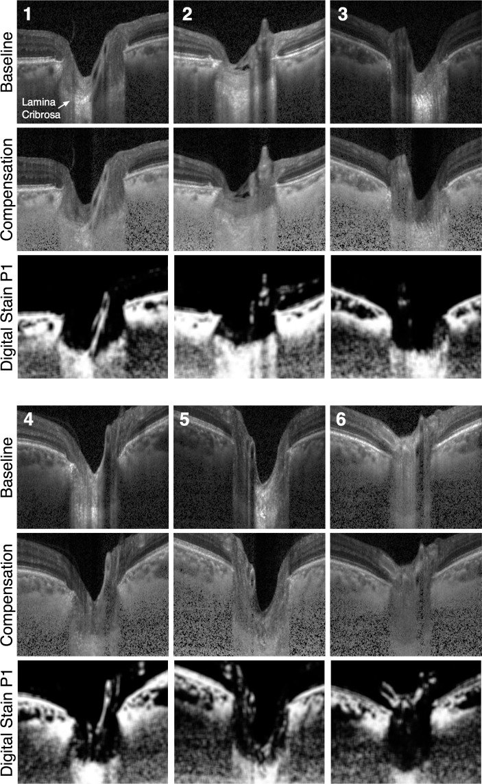
Figure 3.
Baseline, compensated, and digitally stained images (projections P1) for 6 (of 10) healthy ONHs. P1 consistently stained for connective tissues (mostly sclera, choroid, and lamina cribrosa). In the P1 images, the lamina cribrosa was even stained in the nasal region even though strong blood vessel shadowing was observed in the baseline images.

