Figure 1.
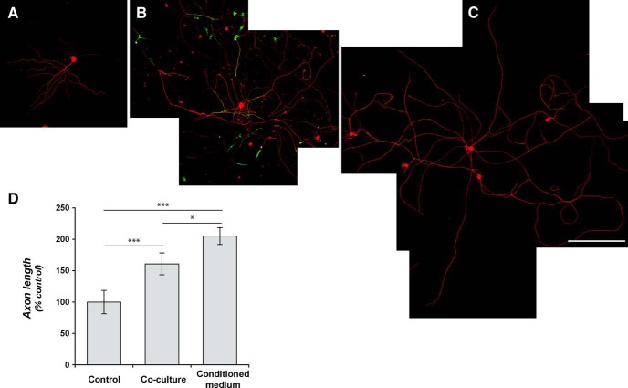
GRPs enhance axonal growth in DRGs in vitro. A–C, Representative montage images are shown for control dissociated DRG culture (A), dissociated DRGs cocultured with rat GRPs (B), and dissociated DRGs cultures exposed to conditioned medium from alkaline phosphatase-expressing rat GRPs (C). βIII-tubulin (red) immunofluorescence highlights the neurons; GRPs are visualized by immunostaining for alkaline phosphatase (green). D shows quantification for the average length of the longest axon per neuron ± SEM (n ≥ 30 neurons in three separate experiments; **p ≤ 0.01 by Student’s t test). Scale bar, 250 μm.

