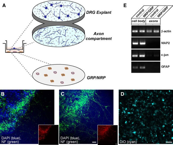
Figure 3.
Approach for DRG/progenitor cell coculture to isolate axonal processes. A, Schematic of modified Boyden chamber used for culture system for isolation of axons from DRG neurons is shown. For this, DRG explants were plated onto the upper surface of a porous PET membrane, as previously used for dissociated DRGs (Zheng et al., 2001), and GRPs/NRPs were cultured on the surface of the plate well. B–D,Representative confocal projection images of upper membrane surface with DRGs (B), corresponding image stacks for lower membrane surface showing a dense array of axons (C), and GRPs/NRPs along the plate surface (D) are shown. DRGs were stained for βIII-tubulin (green) and DAPI (blue). The GRPs were visualized with DiO stain (cyan). The insets in B and C show mCherry signals (red) of AAV5-mChMYR3'amph-transduced DRGs in cell bodies (B) extending into axons along the lower membrane surface (C). Scale bars: B, C, 100 µm; D, 50 µm. E, Representative RT-PCR from cell body and axonal isolates for DRGs cultured under control conditions or over a bed of GRPs/NRPs. The axonal isolates are depleted of cell body (MAP2 and c-Jun) and glial (GFAP) mRNAs, but contain the known axonal transcript β-actin.

