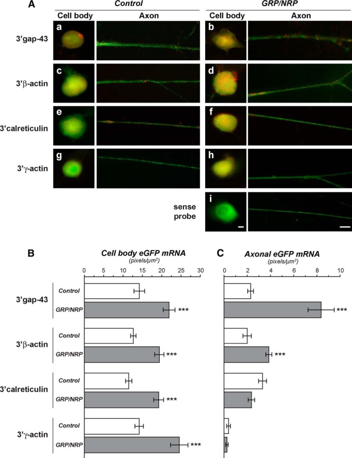
Figure 5.
Alterations in axonal mRNA levels with GRP/NRP coculture is conferred by axonal mRNA UTRs. Aa–i, Representative fluorescent images for eGFP mRNA (red) and βIII-tubulin protein (green) in cell body and distal axon shaft of dissociated DRG cultures transfected with eGFPMYR plasmids with 3'UTRs of rat GAP43 (a, b), β-actin (c, d), CALR (e, f), and γ-actin mRNAs (g, h). i, Images for sense eGFP probe represent the 3'β-actin construct. Left-hand columns of axon and cell body images show control DRG cultures, and right-hand columns show DRGs cocultured with GRPs/NRPs. All image pairs are exposure matched (control vs GRP/NRP coculture axon and control vs GRP/NRP coculture cell body). As previously published, axonal GFP mRNA is not seen for the construct with 3'UTR of γ-actin, but axonal signals are seen for GAP43, calreticulin, and β-actin 3'UTR constructs (Willis et al., 2007; Vuppalanchi et al., 2010; Yoo et al., 2013). B, C, Quantitation of axonal and cell body GFP mRNA intensities across multiple transfection experiments for DRGs with or without GRPs/NRPs are shown as average signal intensities ± SEM (n ≥ 30 processes over three independent transfections and cultures; ***p ≤ 0.001 by Student’s t test). Scale bars, 20 µm.

