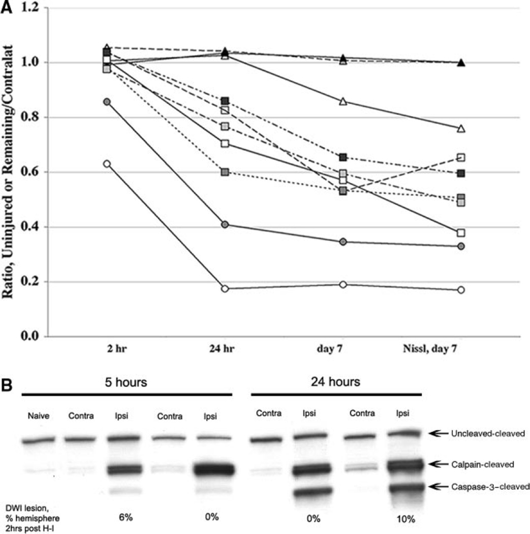
Figure 1.
A, Injury evolution identified by MRI and corresponding histology. MRI was performed at 2 hours, 24 hours, and 7 days after H-I in 10 pups. For the 2- and 24-hour time points, the extent of injury on DWI maps is expressed as the difference between the volume of noninjured ipsilateral hemisphere divided by the volume of the contralateral hemisphere. For the 7-day time point, measurements are done on T2-weighted images and are expressed as the remaining tissue in ipsilateral hemisphere divided by the tissue in the contralateral hemisphere. B, DWI shortly after H-I does not predict the calpain or caspase-3 activation. Calpain activation occurs 5 hours after H-I in animals with no or small (6% of ipsilateral hemisphere) DWI lesions 2 hours after H-I. Both calpain- and caspase-3-dependent spectrin cleavage are seen 24 hours after H-I in animals with no early DWI lesions. Contra, contralateral; ipsi, ipsilateral.

