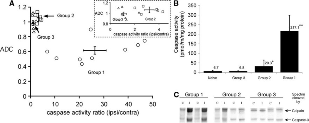
Figure 3.
DWI lesions and caspase-3 activation 24 hours post-H-I. A, ADC of ipsilateral cortical region versus normalized caspase-3 activity (activity in ipsilateral cortex/contralateral cortex). All animals with markedly elevated caspase-3 activity had a reduced ADC value in the ipsilateral cortex. Error bar crosshair shows mean±SD in group 1. Inset, Expanded region for groups 2 and 3. Note that ADC of the cortical tissue was the same for both subgroups, whereas the caspase-3 activity ratio was significantly greater in group 2. B, Caspase-3 activity in brain homogenates from animals in groups 1 to 3. Mean±SD; n=6 to 9 per group. *P<0.05; **P<0.01 in comparison to uninjured. C, In animals imaged 24 hours after H-I, a substantial caspase-3-dependent spectrin cleavage occur in group 1, whereas subtle to no cleavage is seen in groups 2 and 3, respectively. C, contralateral; I, ipsilateral.

