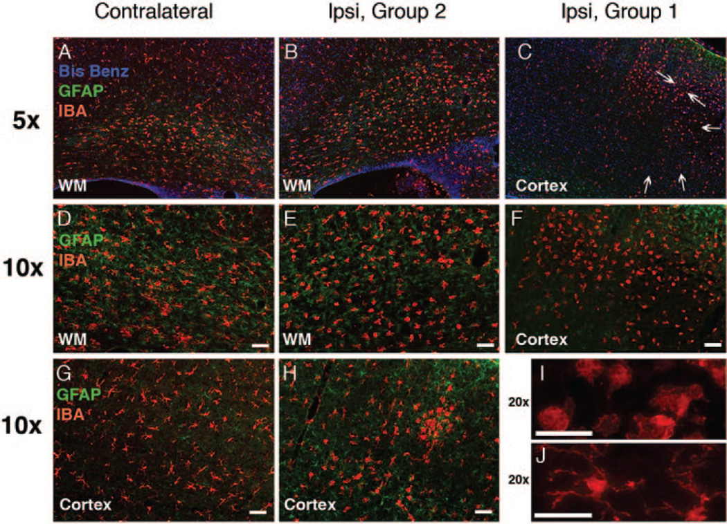
Figure 5.
Microglial activation and reactive astrogliosis associated with profound and modest caspase-3 activation 24 hours after H-I. A–J, Compared with the contralateral hemisphere (A, D, G), a range of microglial morphologies (Iba1, red) is seen in the hemisphere ipsilateral to modest H-I injury (group 2, B, E, H). Accumulation of activated Iba1-positive cells is seen in group 1 (C, F, I). Note a major difference in morphological transformation of microglial cells in group 2 compared with group 1. I–J, Higher magnification picture (20× objective) of activated (I) and resting (J) microglia. Arrows in C indicate substantial damage (morphology of Bis-Benzamide). Modest reactive astrocytosis in the white matter of the H-I hemisphere, group 2 (B, E, glial fibrillary acidic protein, green). In group 1, reactive astrogliosis occurs predominantly outside of severely injured regions (C, F).

