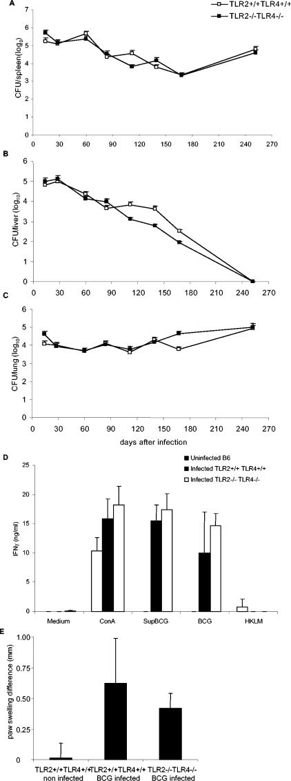
FIG. 8.
Control of BCG infection and T-cell priming in TLR2-TLR4-deficient mice. Viable bacterial load was assessed in spleen (A), liver (B), and lung (C) homogenates of TLR2-TLR4-deficient (▪) and control (□) mice infected with BCG (2 × 10⁶ CFU/mouse, given i.v.). The data are expressed as mean CFU values per whole organ ± the SD (n = four mice per point; P > 0.05). (D) Splenocytes isolated from BCG-infected control and TLR2-TLR4-deficient mice 14 days after infection (2 × 10⁶ CFU/mouse, given i.v.) or from uninfected control mice were restimulated in vitro in the presence of concanavalin A (ConA; 2.5 μg/ml), soluble BCG antigens (Sup BCG, 100 μg/ml), live BCG (two bacteria/cell), or an unrelated antigen, HKLM (100 bacteria/cell). IFN-γ production was quantified in the supernatants after 48 h of incubation. The results are means ± the SD (from n = four mice per genotype). (E) Cutaneous DTH response was performed 8 months after infection of TLR2-TLR4-deficient and control mice with BCG (2 × 10⁶ CFU, given i.v.) by assessing the footpad swelling in response to PPD injection. Paw swelling is defined as the difference in thickness between the left paw injected with PPD and the right paw injected with saline. DTH responses of noninfected TLR2-TLR4 wild-type mice are shown as controls. The data are expressed as means ± the SD (n = 3).

