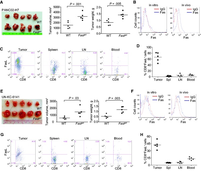
Figure 6.
Role of the Fas-FasL system in suppression of pancreatic tumor growth in vivo. A) PANC02-H7 cells were surgically transplanted to the pancreas of wild-type (WT) C57BL/6 (n = 5) and faslgld (n = 5) mice. Shown are the established pancreatic tumors 15 days after tumor transplant. The tumor volume and tumor weight were analyzed by two-sided t test and are presented in the right panels. B) The cultured PANC02-H7 cells and PANC02-H7 tumor tissues from WT mice were analyzed for cell surface Fas protein level by flow cytometry. C) Cells were prepared from the indicated tissues from tumor-bearing WT mice as shown in (A), stained with 7-AAD, CD8-, and FasL-specific monoclonal antibodies (MAbs), and analyzed by flow cytometry. The 7-AAD-CD8+ cells were gated and quantified for FasL+ cells. D) Quantification of % CD8+FasL+ cells as shown in (C). E) UN-KC-6141 cells were surgically transplanted to the pancreas of WT C57BL/6 (n = 5) and faslgld (n = 5) mice. Shown are the established tumors 15 days after tumor transplant. The tumor volume and tumor weight were analyzed by two-sided t test and are presented in the right panels. F) The cultured UN-KC-6141 cells and UN-KC-6141 tumor tissues from WT mice were analyzed for cell surface Fas protein level by flow cytometry. G) Cells were prepared from the indicated tissues from tumor-bearing WT mice as shown in (E), stained with 7-AAD, CD8-, and FasL-specific MAbs and analyzed by flow cytometry. The 7-AAD-CD8+ cells were gated and quantified for FasL+ cells. H) Quantification of % CD8+FasL+ cells as shown in (G). LN = lymph node; WT = wild-type.

