Figure 1.
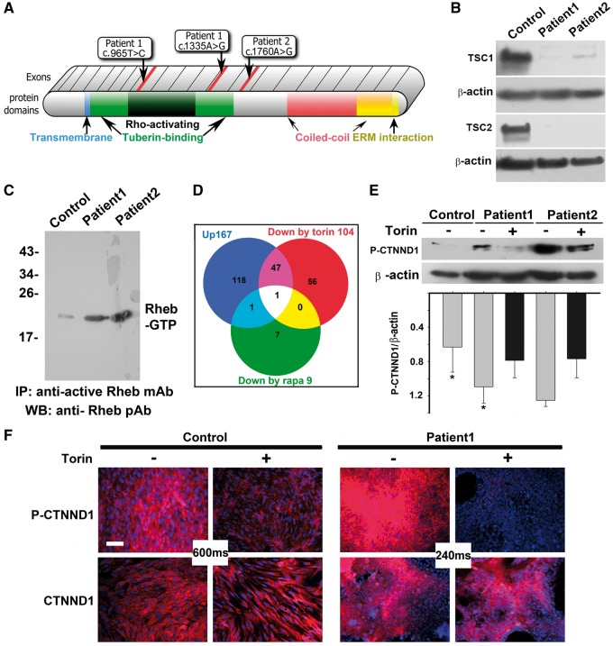
mTOR inhibitors block hyperphosphorylation of CTNND1 serine 268 in TSC patient astrocytes. (A) Diagram of TSC1 protein, showing known motifs and the location of patient mutations. Patient 1 had compound, heterozygous base substitutions in both TSC1 alleles: c.965T > C in the Rho-activating domain of one allele, and C.1335A > G in the tuberin-binding domain of the other allele. Patient 2 exhibited a single c.1760A > G mutation in one of their TSC1 alleles. No deletions were found in microarray analyses. (B) Immunoblot analysis of astrocytic cell lines showed loss of TSC1 or TSC2 protein. Control human astrocytes were from a commercial source (ScienCell Research Laboratories #1800, Carlsbad, CA). (C) Active GTP-bound Rheb is upregulated in TSC patient astrocytes. Lysates from control human astrocytes and patient astrocytes, cultured to 80 to 90% confluence, were immunoprecipitated with an antibody that specifically recognizes GTP-bound Rheb. Immunoprecipitated protein was compared via immunoblot. (D) Stable Isotope Labelling by Amino acids in Cell culture (SILAC) in the absence and presence of Torin1 or rapamycin identified hyperphosphorylation of catenin delta-1 (CTNND1) alone as driving TSC1 mTOR-opathy. Venn diagram depicts proteins hyperphosphorylated in TSC patient astrocyte cultures grown in the presence of vehicle (blue circle; 167 phospo-peptides), rapamycin (green circle; 9 phospo-peptides) or Torin1 (red circle; 104 phospo-peptides). In lysates from TSC astrocytes, both mTOR inhibitors prevented hyperphosphorylation of the mTOR pathway component, CTNND1, on serine 268. (E,F) CTNND1 serine 268 is hyperphosphorylated via the overactive mTOR pathway in TSC patient astrocytes. Confluent cultures of control and patient astrocytes were incubated for 1 h in the absence or presence of the mTOR inhibitor, Torin1. E,Immunoblots of lysates derived from two patient cell lines (normalized to beta actin expression) show hyperphosphorylation of CTNND1 S268 was blocked by Torin1. Band intensity was measured via densitometry and normalized to β-actin levels. The relative value of normal was 0.52 ±0.24, whereas patient values were 1.17 ± 0.3131 (n = 3 immunoblots; *P = 0.048). F, Immunostaining for CTNND1 P-S268 shows hyperphosphorylation (but not expression) of CTNND1 in TSC patient astrocytes is downregulated by Torin1. Image acquisition times were dramatically reduced for mutant cultures (240 ms vs. 600 ms). Scale bar, 100 µm.

