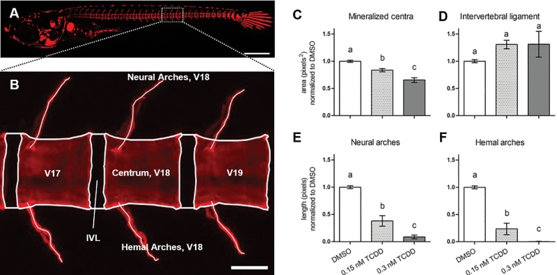
FIG. 2.
Morphological assessment of axial vertebral elements from V17 to 19) of DMSO-treated medaka at 20 dpf. Depicted in (A) is a representative whole-mount individual stained with ALC, 6× magnification. Panel B shows boxed region in A) with morphological atlas labeling V17–19, and centrum, IVL, and hemal and neural arches of vertebra 18 as a representative example, 20× magnification. Centra and IVL areas are shown in (C) and (D), respectively; lengths of neural and hemal arches are shown in (E) and (F), respectively. Values represent the mean from 3 separate measurements taken from V17–19. Letters denote statistical significance between groups analyzed by 1-way ANOVA with Tukey’s post hoc analysis. Scale bars in (A) and (B) denote 500 and 50 µM, respectively.

