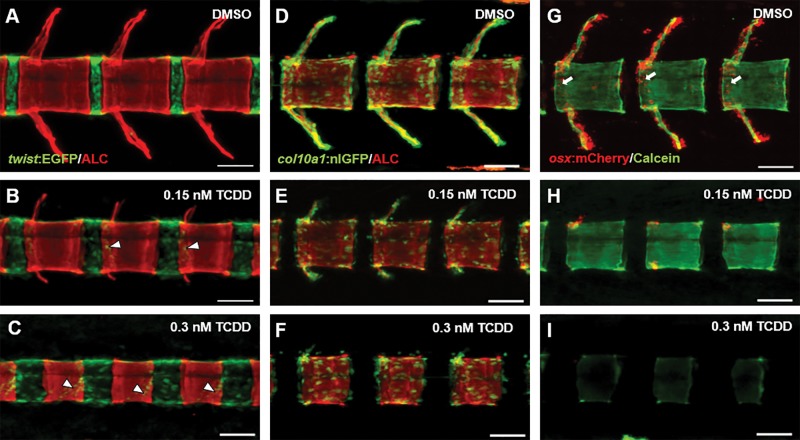
FIG. 3.
Confocal microscopy of representative DMSO- and TCDD-exposed tg(twist:EGFP) (A–C), tg(col10a1:nlGFP) (D–F), tg(osx/sp7:mCherry) (G–I) medaka at 20 dpf. Individuals were stained with ALC or calcein to identify mineralized bone matrix within the context of osteoblasts and osteoblast precursors. Arrowheads in (B) and (C) highlight twist:EGFP+ cells on the mineralized chordacentra. Arrows in (G) denote osx/sp7:mCherry+ cells localized to the periphery of centra undergoing perichordal ossification. Images of V17–19 were captured using the Zeiss LSM 710 confocal system, 20× magnification (scale bars = 50 µM).

