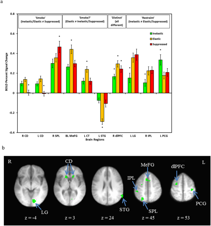
Figure 2. BOLD signal associated with differential activity during the Consider epoch of the CPT.
Panel a presents individual regions organized by common patterns of activation (e.g., ‘Smoke’ reflects greater activation during Inelastic and Elastic demand compared to Suppressed demand). * = choice type is significantly different than the other two choice types. Panel b presents axial slices depicting the locations of the differentially active brain regions, with the exception of the cerebellar tonsil. Radiological conventions are used and side of brain is indicated by R or L (right, left). CD = caudate; SPL = superior parietal lobule; MeFG = medial frontal gyrus; CT = cerebellar tonsil; STG = superior temporal gyrus; dlPFC = dorsolateral prefrontal cortex; LG = lingual gyrus; IPL = inferior parietal lobule; PCG = postcentral gyrus.

