Figure 1.
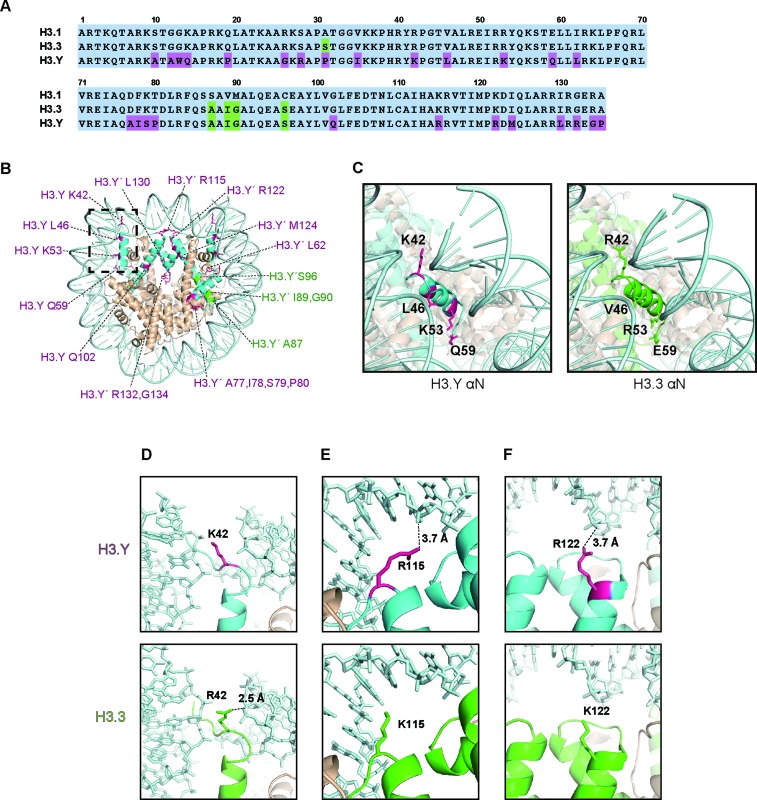
Structure of the human H3.Y nucleosome. (A) Amino acid sequence alignment of the human H3.1, H3.3 and H3.Y proteins. The H3.Y-specific residues are colored purple, and the residues conserved between H3.3 and H3.Y are colored green. (B) Crystal structure of the human H3.Y nucleosome. The H3.Y molecules are colored light blue. The H3.Y-specific residues are represented by purple letters, and the residues conserved between H3.3 and H3.Y are represented by green letters. (C) Close-up views of the αN regions of the H3.Y and H3.3 nucleosomes. The H3.Y αN region encircled by the dotted square in panel B is enlarged and presented with a modified angle (left panel). The H3.Y-specific residues corresponding to K42, L46, K53 and Q59, which are located in the αN region, are colored purple. The H3.3 (green) αN region is also presented with the same angle as in the left panel (right panel) (PDB ID: 3AV2). (D, E and F) Close-up views of the H3.Y-specific (D) Lys42, (E) Arg115 and (F) Arg122 residues (left panels), corresponding to the H3.3 Arg42, Lys115 and Lys122 residues (middle panels). The right panels show the merged views of these residues of the H3.Y and H3.3 nucleosomes.

