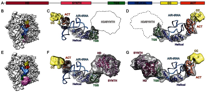
Figure 3.
RelA adopts an extended conformation on the ribosome. (A) Schematic showing the domain organization of E. coli RelA with HD (magenta), SYNTH (pink), TGS (green), HELICAL (blue), CC (yellow) and ACT (orange) subdomains. (B) Overview of RelA-SRC with subdomains colored according to (A) and A/R-tRNA (light blue). (C and D) Complementary views showing isolated electron density of RelA with fitted homology models for TGS (green, PDB2EKI) and ACT (orange, PDB2KO1) subdomains, as well as model poly-Alanine helices fitted to the helical linker region (blue) and density for CC colored in yellow. (E–G) As (B–D) with 12 Å filtered maps and additional fitted homology models for HD (magenta) and SYNTH (pink) based on PDB1VJ7 (24).

