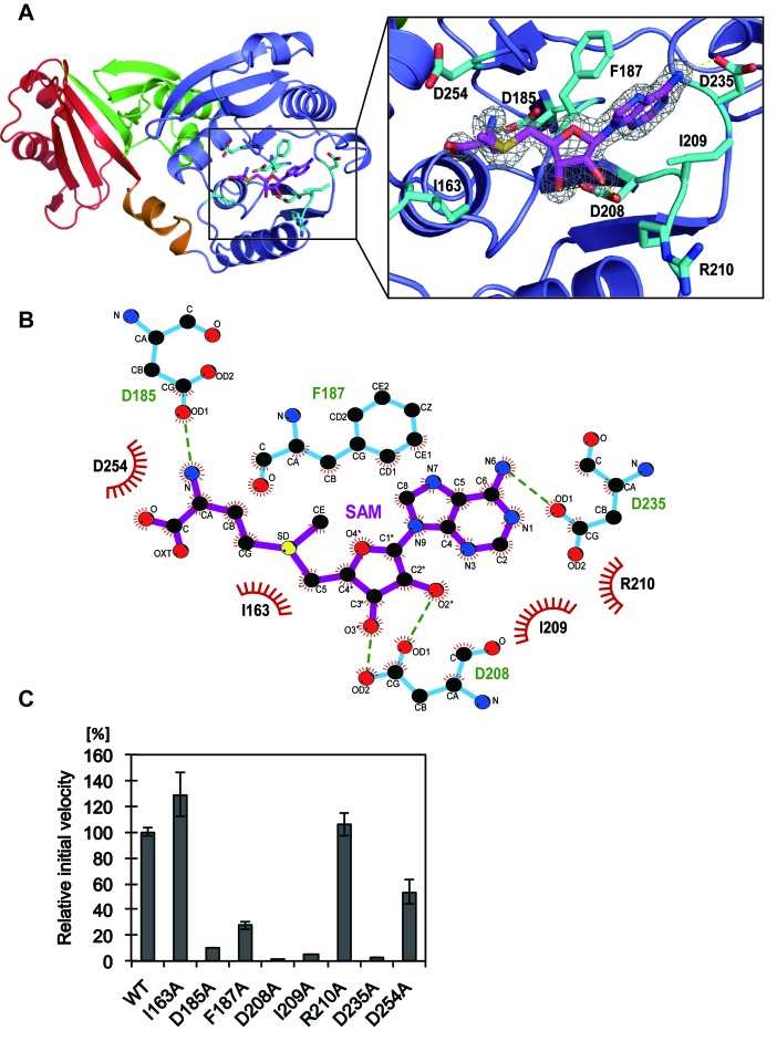
Figure 3.
The SAM-binding site of TkoTrm11 in complex with SAM. (A) Close-up view of the SAM molecule and its binding residues illustrated as stick models (magenta and cyan, respectively). Electron density map around the SAM molecule contoured at 2.0 σ. (B) Ligplot diagram (64) of interactions between TkoTrm11 amino acid residues and the bound SAM molecule. (C) Methyl transfer activity of the alanine-substituted mutant proteins. The mutant name indicates the site of alanine substitution. The initial velocity of wild-type (WT) TkoTrm11 is expressed as 100%. Error-bar indicates the SD (standard deviation) that was calculated between three independent experiments.

