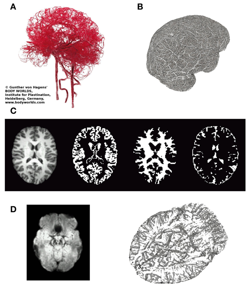
Figure 2. Illustrates relevant macroscopic components in BOLD-based fMRI of the brain.
(A) Arterial vessels ex vivo (copyright Gunther von Hagens, Körperwelten, Institut für Plastination, Heidelberg, www.koerperwelten.de), (B) MR-venography of venous vessels in vivo at 7 Tesla [courtesy Dr. M. Barth; adapted from Koopmans [36]]. (C) Representative slice across the brain of a young healthy subject. T1-weighted structural image, segmented gray matter mask, segmented deep white matter mask and segmented CSF space (from left to right). Note that in contrast to (A,B), vessels are almost invisible. (D) Mean SWI (n = 3, left), highlighting brain regions with strong susceptibility differences (dark regions) causing artifacts (e.g., frontal lobe/nasal cavities, temporal lobe/ear canals, veins near the brain stem, etc.). MR-venogram (n = 1), visualizing the basal vein of Rosenthal, running next to the amygdalae and brainstem.

