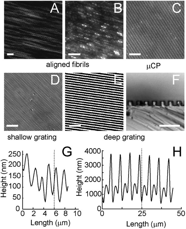
Figure 1. Length Dimensions of different substrates.

(A) Epitaxially grown, aligned collagen fibril image taken using atomic force microscopy. Calibration bar length = 200 nm. (B) aligned collagen fibrils binding carboxylate-functionalized 40 nm polystyrene fluorescent spheres and imaged under fluorescence microscopy. (C) μCP of alexa 555 collagen. Phase contrast imaging of a (D) shallow grating and (E, F) deep grating taken through the substrate (D, E) and of a slice of substrate laid on its edge (F). Calibration bar length = 30 μm (D, E) or 5 μm (F).
