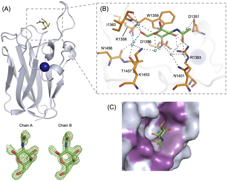
Fig 6. GalNAc binding determinants of CpGH31 CBM32-2.
(A) Backbone cartoon representation of CBM32-2 (grey) in complex with GalNAc (green), solved to a resolution of 2.00 Å. The associated calcium ion is depicted as a blue sphere. Fobs-Fcalc electron density maps of GalNAc (green) bound to peptide chains A and B of the CBM32-2:GalNAc structure are shown in green mesh and contoured to 3.0 σ. (B) GalNAc (green) is bound to CBM32-2 by a several aromatic and polar residues (orange) via direct and water-mediated hydrogen bonds. Associated water molecules are shown as cyan spheres and hydrogen bonds are depicted by dashed lines. The stacking interaction is mediated by Trp1359. (C) The shallow GalNAc-specific binding site of CBM32-2 (shown in grey) accommodates the O4 hydroxyl group of the ligand in an axial position only. The sugar associates with the side chain of Trp1359 (light purple) and forms numerous hydrogen-bonding interactions (magenta) that target the O6 hydroxyl and 2-acetamido groups on either end of the sugar.

