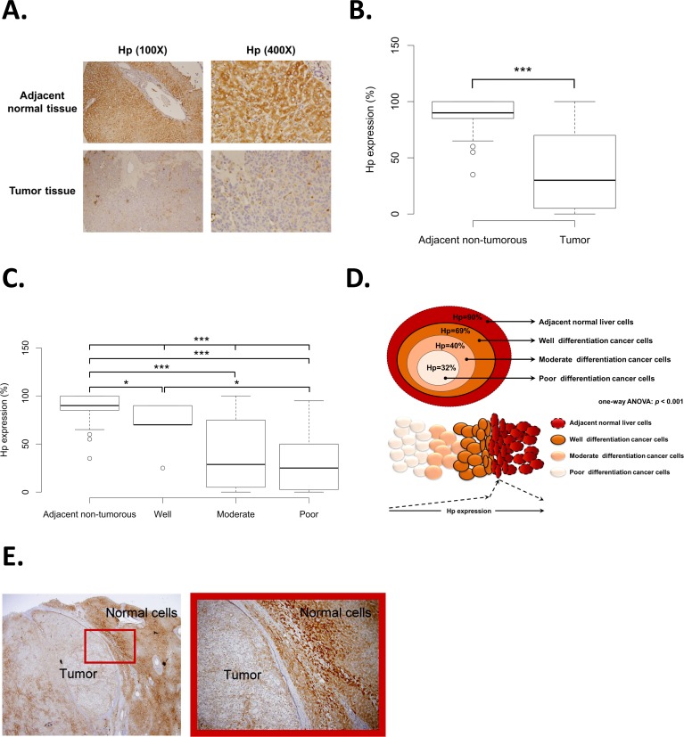
Fig 1. Hp protein expression and cancer differentiation analysis of CCH HCC patients.
(A) An example of Hp IHC staining in adjacent non-tumorous tissues and tumor tissues under 100X and 400X magnification. (B) Hp protein expression analysis between matched pair adjacent non-tumorous tissues and tumor tissues of HCC patients from CCH (p < 0.001). (C) Boxplot of Hp protein expression in adjacent non-tumorous tissues and three stages HCC cancer differentiation (well, moderate, and poor) (p < 0.001). (D) The schematic diagram interpreted the correlation between Hp expression and HCC cancer differentiation (p < 0.001). (E) An example of IHC staining for measuring the Hp protein expression at the junction area between adjacent non-tumorous tissue and tumor tissue.

