FIG. 2.
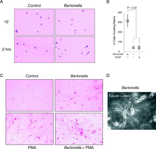
Bartonella promotes invasion, survival, and differentiation in 3-D collagen gels. Infected HUVEC were embedded in type I collagen gel. Where indicated, VEGF (10 ng/ml) or PMA (16 nM) was maintained throughout the assay. (A) Endothelial morphology at 15 min and 2 h after collagen gel formation (H&E staining; magnification, ×400). Note, the elongated shape of infected HUVEC at 2 h. Some HUVEC are cut in cross section, and their elongated shape therefore cannot be appreciated. (B) Numbers of invading (elongated) cells at 2 h (medians and ranges from four separate assays, with one field [magnification, ×100] counted from each). (C) Endothelial morphology at day 4 (H&E staining; magnification, ×100). (D) Tubule from Bartonella infection assay at day 4 (phase contrast; magnification, ×400). The arrow points to an interior lumen running the length of a branching tubule. Parts of the tubule are outside the plane of focus, demonstrating its three-dimensional nature.

