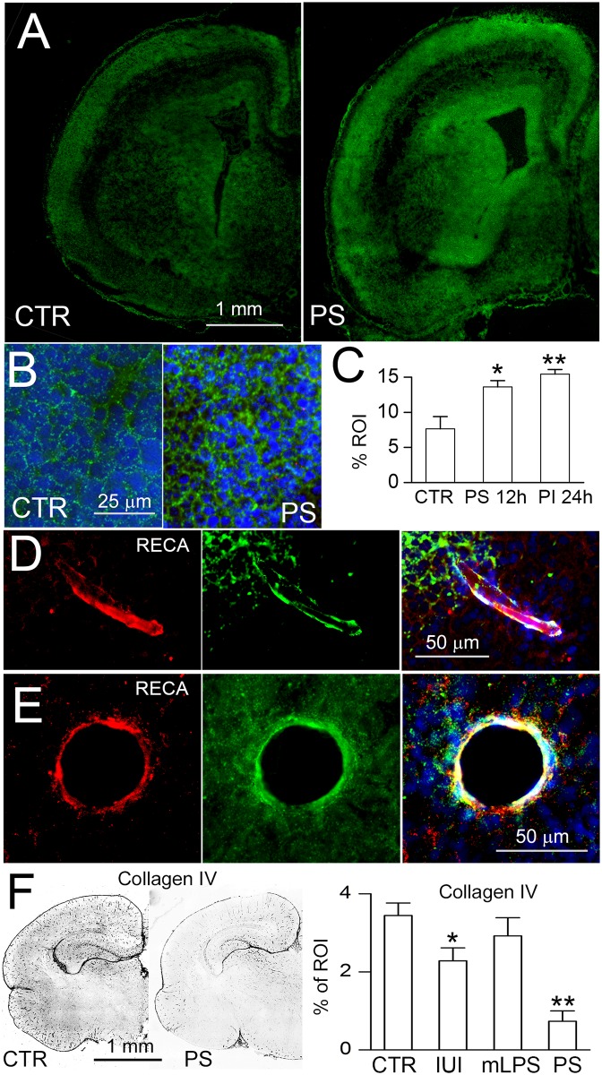
Fig 2. IUI+mLPS increases proteolytic activity and decreases collagen IV immunoreactivity.
A–C: In situ zymography (A,B), with quantification (C), of coronal sections from naïve control (CTR) and 24 hours after dual prenatal pro-angiogenic stimuli (PS) of IUI+mLPS, shown at low (A) and at high (B) magnification; the subventricular zone is shown in (B); nuclei stained with DAPI (blue); scale bars, 1 mm (A), 25 μm (B); 3 pups per group; *, p<0.05; **, p<0.01. D,E: Images of vessels identified by immunolabeling for RECA (red), that show proteolytic activity on in situ zymography (green); merged images are shown on the right; nuclei stained with DAPI (blue); scale bars, 50 μm. F: Immunolabeling for collagen IV (left), with quantification (right), on P0 in naïve controls (CTR), after IUI alone, after mLPS alone, and after the dual pro-angiogenic stimuli of IUI+mLPS (PS), in all cases after vaginal delivery, in coronal brain sections; 5 pups per group; tissues from the IUI alone group were from a previous study [20].

