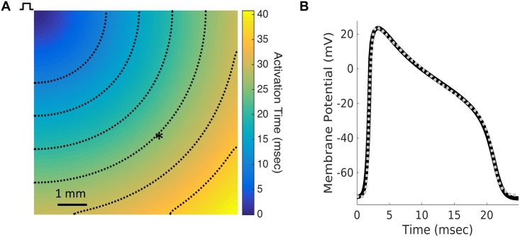
Fig 2. Model action potential replicates experimental action potential.
(A) Model fitting was performed by simulating conduction in a 2-D monolayer and recording an action potential 6 mm from the stimulus site (asterisk). Dashed lines are isochrones of activation at intervals of 5 ms. (B) The action potential generated by the fitted Ex293 membrane model (solid black line) replicates the morphology of the experimentally-recorded Ex293 action potential from [7] (dashed gray line).

