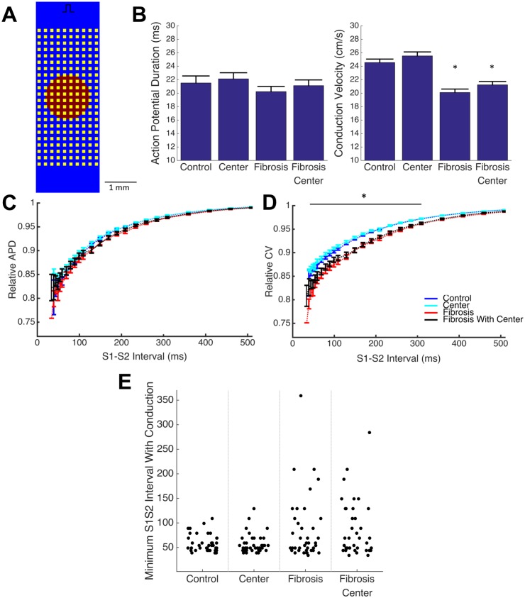
Fig 8. Spatial organization of ionic variation does not affect macroscopic conduction.
The introduction of a central region with reduced variance and prolonged APD (A, red) into a tissue model with and without non-conductive fibrosis-like obstacles (A. yellow) does not cause additional conduction slowing and APD shortening at 1 Hz pacing beyond the effect of fibrosis alone (B). A fibrosis induced exaggeration of CV slowing (D), but not APD shortening (C), at short diastolic (S1-S2) intervals (plotted as mean +/- standard error) is also unaffected by the spatial organization of variation. In addition, spatial organization maintains but does not enhance premature failure, as characterized by minimum S1-S2 intervals able to fully conduct across the domain (E). (* p < 0.05 main effect of fibrosis)

