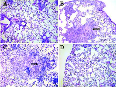
FIG. 4.
Histopathological examination of lung tissue (stained with hematoxylin and eosin) from mice 28 days after challenge with M. tuberculosis. Lung sections from mice vaccinated with HPLC-purified HBHA (A), with only the DDA-MPL adjuvant (B), with recombinant His-tagged HBHA purified from M. smegmatis (C), and with BCG vaccine (D) are compared. The arrows indicate areas of dense cellular infiltration and consolidation typical of tuberculosis in mice at 30 days. Magnification, ×100.

