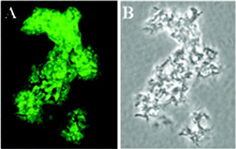
FIG. 5.
Recognition of cell surface antigen on the M. tuberculosis Erdman strain by IgG in sera from mice immunized with HPLC-purified HBHA. Paraformaldehyde-treated bacteria were incubated with pooled mouse sera at a dilution of 1:250, followed by fluorescein isothiocyanate-conjugated anti-mouse IgG. Uniform fluorescence was observed for most bacteria (A) found in a colony of M. tuberculosis visualized by phase-contrast microscopy (B). Fluorescence was visualized by using a Nikon Optiphot-2 microscope with a ×100 phase/fluorescent objective; images were photographed with a SPOT RT digital camera, and the composite was produced by using Adobe Photoshop. Magnification, ×1,000.

