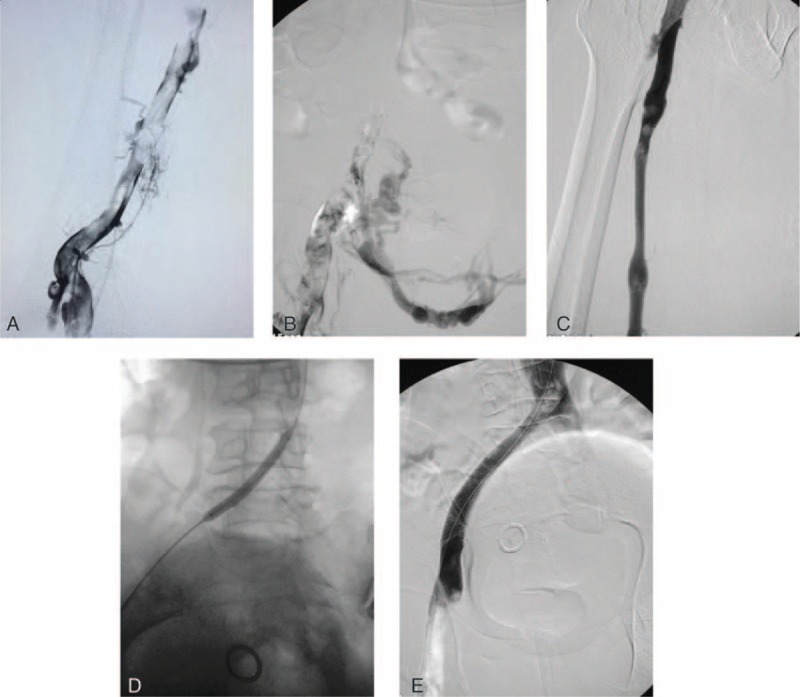
FIGURE 1.

A, Initial venography in the prone position shows femoral vein thrombosis. B, A venography shows occlusion of the common iliac vein and collateral formation. C, The venography shows a patent common femoral vein and little residual thrombus was shown after manual aspiration thrombectomy. D, The common iliac vein was dilated with an angioplasty balloon. E, A venography after stent deployment shows a patent common iliac vein, good antegrade flow, and abolition of collaterals.
