
FIGURE 5.
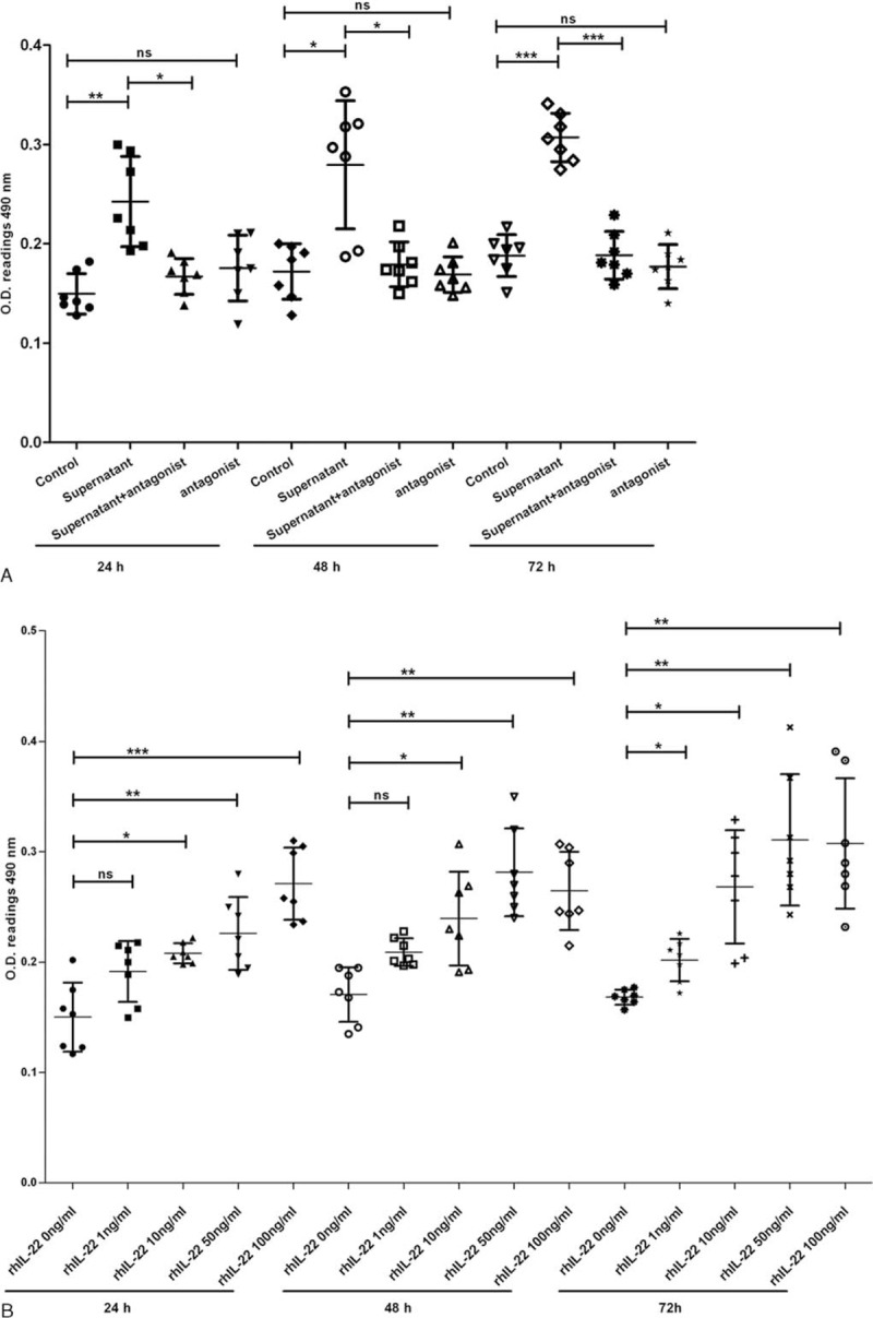
NKp44+NK cells promote the proliferation of FLS via IL-22. The FLS were treated by 50% NKp44+NK cell culture supernatants, 50 μg/mL IL-22 antagonist, combination of both of them, different concentrations of rhIL-22, and negative controls for 24, 48, or 72 hours, respectively. They were then analyzed by MTT assay. NKp44+NK cells culture supernatant promote the proliferation of FLS (A). IL-22 antagonist blocks those promotion process (A). Different concentrations of rhIL-22 promote the proliferation of RA-FLS (B). Values represent mean ± SD of O.D. 490 nm. ∗P < 0.05; ∗∗P < 0.01; ∗∗∗P < 0.001; ns, no significant difference. The data are representative of seven independent experiments. FLS = fibroblast-like synoviocytes, IL-22 = interleukin 22, MTT = methyl thiazolyl tetrazolium, NK = natural killer, RA = rheumatoid arthritis, rhIL-22 = recombinant human interleukin 22, SD = standard deviation.