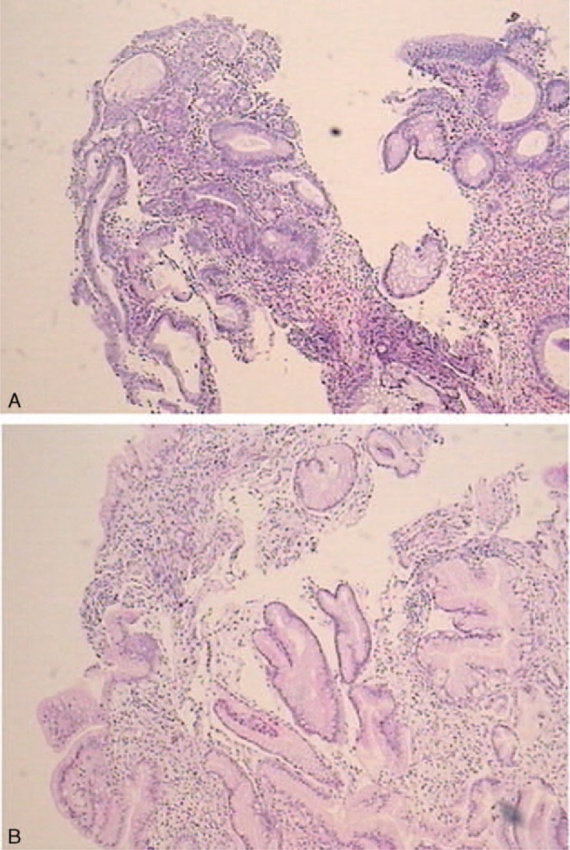
FIGURE 3.

(A) Biopsies from polyps in the antrum showing chronic inflammation in mucosa, stromal hyperemia, and edema, and focal hyperplasia. (B) Biopsies from polyps in the colon showing a mixture of inflammatory cells consisting of lymphocytes and neutrophil granulocytes and clusters of epithelioid cells, crypt abscesses, stromal edema and hyperemia, infiltration of eosinophile granulocytes, and focal hyperplasia of epithelial cells.
