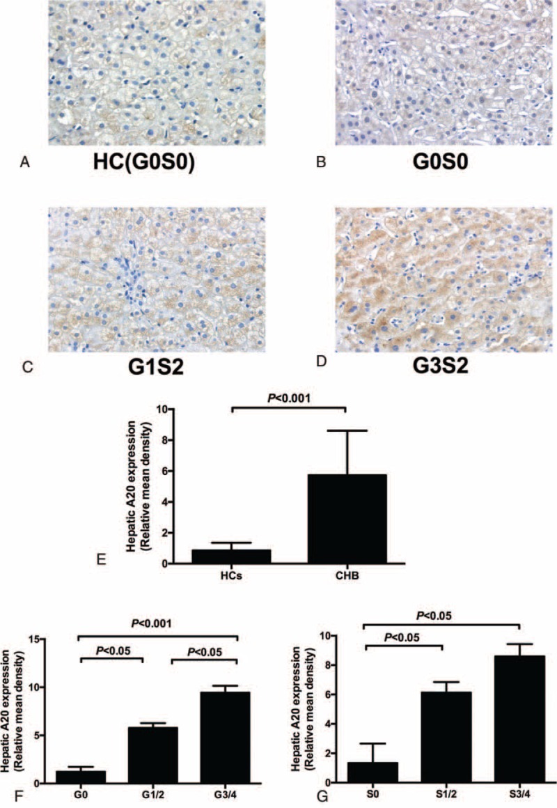
FIGURE 3.

Expression of intrahepatic A20 protein in liver tissues using the immunohistochemical method. A20 positivity was found nearly in all hepatocytes (B,C,D) (200×). The intensity of staining of A20 was correlated with the severity of liver inflammation (B,C,D) (200×). Relative mean density analysis showed the difference in hepatic A20 staining between the CHB group and healthy controls (E). (F) The difference in hepatic A20 staining between inflammation grade (1–2) and grade (3–4). (G) Hepatic A20 staining in fibrosis stage (3–4), stage (1–2), and fibrosis stage 0. CHB = chronic hepatitis B.
