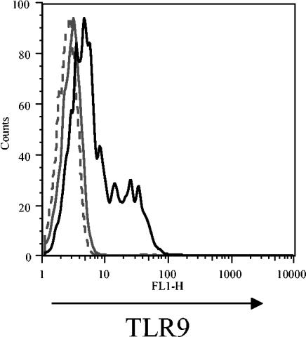
FIG. 5.
Specificity of TLR9 staining. The specificity of the TLR9 staining was further tested by assessing cell surface TLR9 expression on RAW 264.7 cells that were mock transfected or transfected with a plasmid containing human TLR9. At 24 h posttransfection, cells were stained with mouse anti-human TLR9 antibody, which was detected with an FITC-goat anti-mouse IgG/IgM antibody. The histogram plot shown depicts mock-transfected cells stained with only the secondary reagent (gray dashed line), mock-transfected cells stained with the TLR9 antibody plus the secondary detection reagent (gray solid line), and TLR9-transfected RAW cells stained with the TLR9 antibody plus secondary detection reagent (black solid line). RAW cells transfected with human TLR9 display cell surface TLR9 staining distinct from that of the mock-transfected cells. The data shown are from one experiment and are representative of those from three independent experiments.

