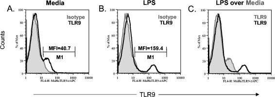
FIG. 6.
Cell surface TLR9 expression on human PBMC is up-regulated following LPS stimulation. Human PBMC were cultured for 24 h in 30, 10, or 1 μg of LPS per ml and then stained with the murine anti-human TLR9-APC MAb or the isotype control. The histogram plots shown are (A) PBMC cultured in medium alone stained with the TLR9 antibody (black line) overlaying the isotype control (gray histogram), (B) human PBMC cultured in LPS (10 μg/ml) stained with the TLR9 antibody (black line) overlaying the isotype control (gray histogram), and (C) human PBMC cultured in LPS (10 μg/ml) stained with the TLR9 antibody (black line) overlaying human PBMC cultured in medium alone stained with the TLR9 antibody (gray histogram). The MFIs for the TLR9+ cells are shown in histograms (A) and (B). No difference in the MFI of TLR9 up-regulation was observed among the various concentrations of LPS (data not shown). The data shown are representative of those from three independent experiments.

