FIG. 7.
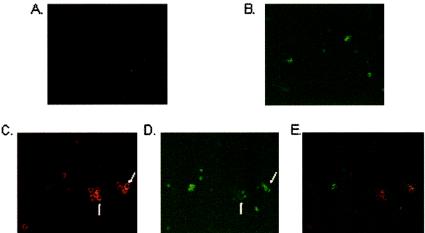
Cell surface TLR9 expression visualized by immunofluorescence staining. Double staining of LPS-stimulated human PBMC with (A) the APC-labeled isotype-matched MAbs and (B) FITC-labeled anti-CD19 MAbs, using the same microscopic field, and double staining of LPS-stimulated PBMC with (C) the APC-labeled anti-TLR9 MAbs and (D) FITC-labeled anti-CD19 MAbs, using the same microscopic field, are shown. Staining was observed under a fluorescence microscope at a magnification of ×40. Arrows indicate cell surface TLR9 staining (C), which did not colocalize with CD19 staining (arrows in panel D). (E) Staining observed with the anti-CD19 MAbs (Fig. 6D) overlaid by the staining observed with the TLR9 MAbs (Fig. 6C) shows distinct cells expressing TLR9 (red) and CD19 (green).

