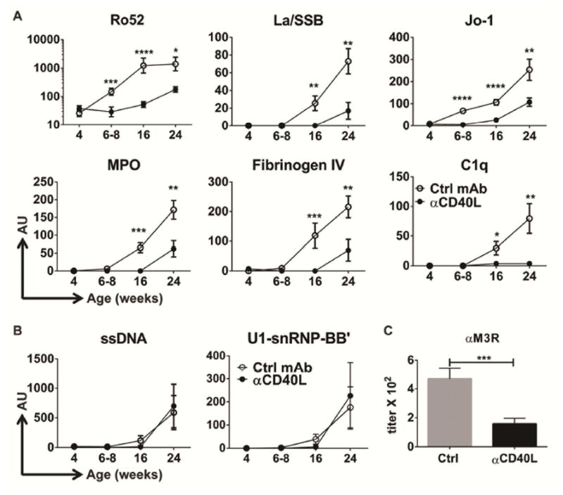
Figure 5. Anti-CD40L Ab administration to young animals prevents autoantibody development of defined specificities.

(A–B) Female NOD.H-2h4 mice received a single injection of either isotype control (Ctrl, white circles) or anti-CD40L mAb (black circles) at 4 weeks-of-age, and serum was collected serially at the indicated ages and screened for reactivity to self-antigens using a 95-autoantigen array. (A) Examples of IgG autoantibodies significantly decreased by anti-CD40L treatment compared to control. (B) Examples of IgG autoantibodies unaffected by anti-CD40L treatment compared to control. Data represents n= 11 for control mAb-treated mice and n= 13 for anti-CD40L-treated mice from three independent experiments. AU= Mean normalized fluorescence IgG signal ± SEM. * p< 0.05; ** p<0.01, *** p<0.001, **** p<0.0001. (C) End point titer (mean +/− SEM) of anti-M3R IgG autoantibodies in sera from of 20–26 week-old female NOD.H-2h4 mice that received a single treatment of either isotype control antibody (n=12) or anti-CD40L antibody (n=15) at 4 weeks-of-age. Data represents two independent cohorts.
