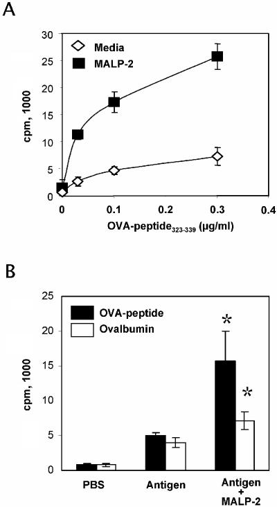
FIG. 6.
Effect of MALP-2 treatment on antigen presentation. (A) OVA-specific CD4+ T cells purified from DO11.10 mice were cocultured with macrophages loaded in vitro with serial dilutions of the OVA peptide in the presence or absence of MALP-2 (0.25 μg/ml). (B) Dendritic cells, which were isolated by magnetic sorting for CD11c+ cells from NALT of mice that received OVA peptide (200 μg) or OVA protein (10 mg), in the presence or absence of MALP-2 (0.5 μg) by the intranasal route. Cells were cocultured during 4 days, and T-cell proliferation was assessed by [3H]thymidine incorporation. Results are expressed as the mean counts per minute values from triplicate wells; standard deviations are indicated by vertical lines. *, statistically significant at a P level of <0.05 by Student's t test.

