
Fig 4. Comparison of simulations to contraction of a cardiomyocyte.
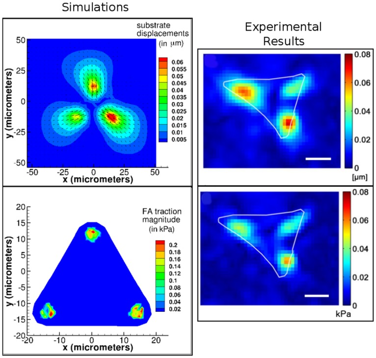
Our simulations are shown in the left panel, and experiments from Hersch et al. [40] are on the right (with permission under CC-BY 4.0 license). Experimental result figures were modified from original by including scale bar and removing letter referring to original figure from top left corner. Scale bar = 10 μm. The substrate displacements from simulations compare well to those obtained experimentally both qualitatively and quantitatively. FA tractions, whose magnitudes are illustrated as contours, compare well qualitatively. Cell and substrate Young’s modulus: Ec = 20 kPa, Es = 15 kPa. FA springs rupture at a stretch of 0.4 μm.