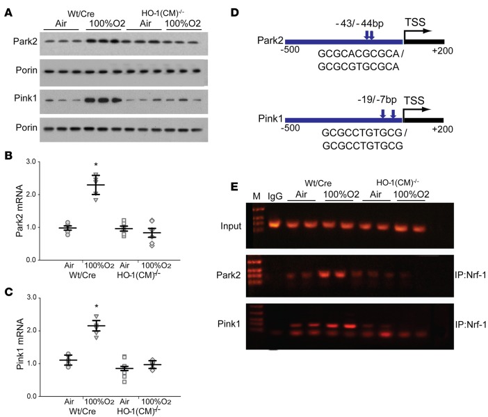
Figure 6. HO-1 is required for Pink1 and Park2 gene expression through NRF-1 activation after hyperoxia.
(A) Representative Western blots of Parkin (Park2) and PINK1 in mitochondrial fractions of the heart from WT/Cre and HO-1(CM)–/– mice prepared before and after hyperoxia. Porin was used as a loading control. (B) Deletion of HO-1 in HO-1(CM)–/– mice decreases cardiac expression of Park2 mRNA. (C) Lack of HO-1 in the heart of HO-1(CM)–/– mice decreased expression of PINK1 mRNA. (D) Schematic diagrams (–500 to +200 bp) of the Pink1 (ENSMUST00000030536) and Park2 (ENSMUST00000191124) promoter regions. Sequences were aligned between human and mouse using rVISTA 2.0. A search for NRF-1 binding sites upstream of the transcription start site (TSS) identified multiple consensus motifs for NRF-1 in human and mouse Pink1 and Park2 gene promoters by Genomatix and DNAsis software. (E) NRF-1 occupancy of promoters in vivo was investigated by ChIP analysis. Input lanes show the PCR product derived from chromatin prior to immunoprecipitation. Antibodies against NRF-1 used for immunoprecipitation, while IgG was used as negative control. Precipitated DNA was analyzed by PCR with primer sets specific for the 3 promoters (mean ± SEM; horizontal bars represent mean values. *P < 0.05 for pre- vs. posthyperoxia; n = 6 per group; 2-way ANOVA).

