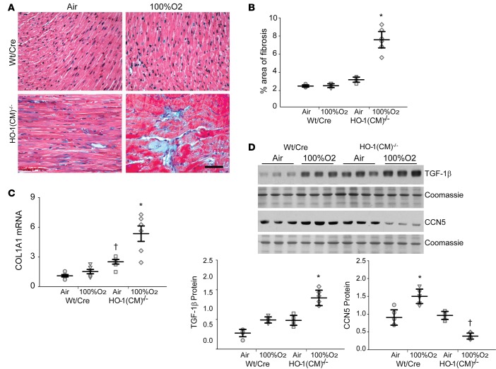
Figure 7. HO-1 deletion enhanced cardiac fibrosis after hyperoxia.
(A) Heart sections were stained with Masson’s trichrome to locate collagen (blue, fibrous collagen), which revealed extensive fibrosis of the left ventricle of HO-1(CM)–/– mice, mainly after hyperoxia (Scale bar = 50 μm). (B) The graph shows quantitation of fibrosis by densitometry. (C) Analysis of Col1A1 mRNA levels in the two strains of mice before and after hyperoxia. (D). Western blots of TGF-β and CCN5 proteins expression in heart lysates prepared before and after hyperoxia. The graph shows TGF-β and CCN5 protein quantification by densitometry relative to coomassie loading control (mean ± SEM; horizontal bars represent mean values; *P < 0.05 for pre- vs. posthyperoxia; †P < 0.05 for WT/Cre vs. HO-1(CM)–/–; n = 6 per group; 2-way ANOVA.).

